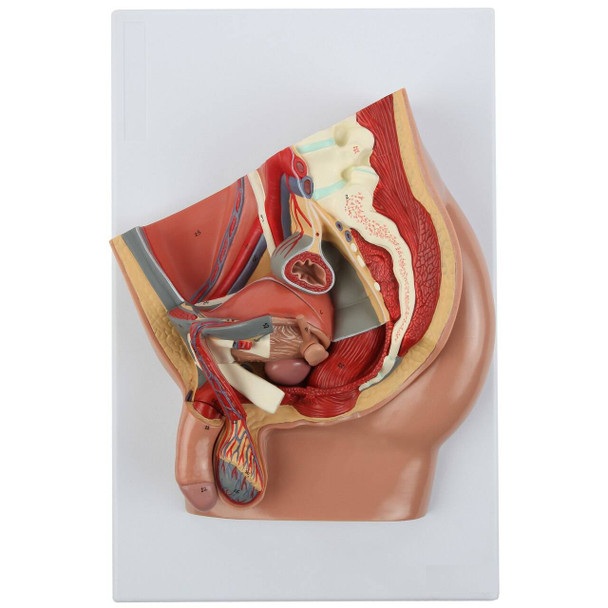
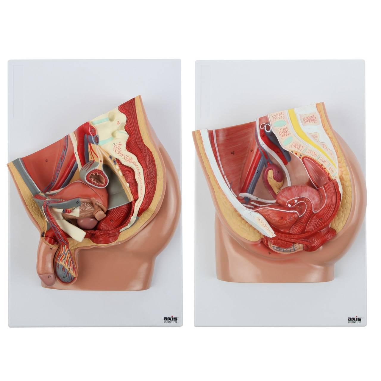
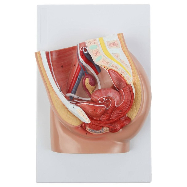
4-Part Median Section Male Pelvis Anatomy Model with Removable Parts
Human Male Pelvis Model is an anatomically accurate representation of the male pelvis anatomy. This model offers a complete view of the male reproductive organs, urinary system, pelvic floor, and lumbar anatomy. This model has removable organs for a more thorough study of each anatomical part.
The pelvis model is mounted on a sturdy base for study, display, and easy transport of the model. The model is numbered and includes a full-color detailed study guide that identifies all 39 anatomical parts on the model. Study the difference in the layers of muscle, organs, systems, and venous drainage of the male pelvis with this highly detailed anatomy model.
FEATURES
Male Pelvis Anatomy Model
Human Male Pelvis Model features the reproductive and urinary organs in the male pelvis. This model includes removable parts, as well as some lumbar anatomy and the various vasculature in the area. Aside from reproductive and urinary organs, some parts of the digestive tract are also featured in the model. The rectum and anus are presented in their correct orientation and placement in relation to the pelvic floor.
The rectum and anus featured on the model is a useful education tool in studying the adjacent organs and structures surrounding them. This mode can be used to determine correct palpations to diagnose diseases. This pelvis model is a perfect visual tool to display distinctions and relations between the different systems and structures in the pelvic cavity.
Reproductive System and Organs
The 4-part male pelvis model features both external and internal male reproductive organs. The external reproductive organs include the penis, testicles, scrotum, and epididymis. The model includes a removable penis with a seminal vesicle for a more specific study of the organs. Once the penis is detached, there is a cross-section display of the remaining external reproductive organs which are rooted in their correct locations in the pelvic cavity to aid in the study of the connection between these organs.
On the internal side, the vas deferens, prostate gland, and spermatic cord are also represented with detail and accuracy. These accessory organs and vessels contribute to the movement of sperms through the male reproductive system. This male pelvis model illustrates the connections between the reproductive organs and the other systems and structures that course through the pelvic cavity.
Urinary System and Organs
The male pelvis model shows the urinary system and its organs. The model includes a removable bladder with the ureter, pubic bone, prostate, and spermatic cord attached. Other details on the removable organ include textures, orifices, rings, arteries, and veins.
The model also features muscle attachment sites in relation to the viscera in the male pelvic cavity, highlighting the abdominal internal oblique (attachment site) and interfoveolar ligament (attachment site) which are adjacent to the bladder. This model is perfect for comparative anatomy regarding male and female pelvic organs, including the differences between the male and female urinary tracts.
Veins and Arteries
The common iliac vein, whose function is to return drained deoxygenated blood back to the heart, is featured prominently on the model. The femoral vein that runs through the thighs and aids in limb function is also represented on the model to display its correct location. The iliac artery, femoral artery, and testicular artery (arteries that supply blood to the lower leg and male genital organs) are also well-represented on the pelvic model.
Base Mount
Our 4-Part Human Male Pelvis Model is mounted on a base to fully display a cross-section view of the pelvic cavity.
Reproductive Organs:
- Penis
- Testicle
- Epididymis
- Spermatic Cord
- Prostate Gland
- Seminal Vesicle
Urinary Organs:
- Ureter
- Bladder
- Urethra (Internal and External Orifice)
- Prostate Gland
Digestive Organs:
- Anus
- Rectum